Hemoglobin-PEG Interactions Probed by Small-Angle X-ray Scattering: Insights for Crystallization and Diagnostics Applications

Autor
Baranova, Iuliia
Angelova, Angelina
Stransky, Jan
Andreasson, Jakob
Angelov, Borislav
Datum vydání
2024Publikováno v
Journal of Physical Chemistry BRočník / Číslo vydání
128 (38)ISBN / ISSN
ISSN: 1520-6106ISBN / ISSN
eISSN: 1520-5207Metadata
Zobrazit celý záznamKolekce
Tato publikace má vydavatelskou verzi s DOI 10.1021/acs.jpcb.4c03003
Abstrakt
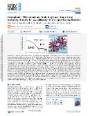
Protein-protein interactions, controlling protein aggregation in the solution phase, are crucial for the formulation of protein therapeutics and the use of proteins in diagnostic applications. Additives in the solution phase are factors that may enhance the protein's conformational stability or induce crystallization. Protein-PEG interactions do not always stabilize the native protein structure. Structural information is needed to validate excipients for protein stabilization in the development of protein therapeutics or use proteins in diagnostic assays. The present study investigates the impact of polyethylene glycol (PEG) molecular weight and concentration on the spatial structure of human hemoglobin (Hb) at neutral pH. Small-angle X-ray scattering (SAXS) in combination with size-exclusion chromatography is employed to characterize the Hb structure in solution without and with the addition of PEG. Our results evidence that human hemoglobin maintains a tetrameric conformation at neutral pH. The dummy atom model, reconstructed from the SAXS data, aligns closely with the known crystallographic structure of methemoglobin (metHb) from the Protein Data Bank. We established that the addition of short-chain PEG600, at concentrations of up to 10% (w/v), acts as a stabilizer for hemoglobin, preserving its spatial structure without significant alterations. By contrast, 5% (w/v) PEG with higher molecular weights of 2000 and 4000 leads to a slight reduction in the maximum particle dimension (D(max)), while the radius of gyration (R(g)) remains essentially unchanged. This implies a reduced hydration shell around the protein due to the dehydrating effect of longer PEG chains. At a concentration of 10% (w/v), PEG2000 interacts with Hb to form a complex that does not distort the protein's spatial configuration. The obtained results provide a deeper understanding of PEG's influence on the Hb structure in solution and broader knowledge regarding protein-PEG interactions.
Klíčová slova
polyethylene-glycol, proteins, state, mechanism, visualization, precipitation, stability
Trvalý odkaz
https://hdl.handle.net/20.500.14178/2953Licence
Licence pro užití plného textu výsledku: Creative Commons Uveďte původ 4.0 International


